At Desoto Dentures and Implants in Southaven, MS, the intraoral camera offers a clear, close-up view of your mouth to support accurate exams and straightforward conversations about care. The intraoral camera helps capture detailed images dentist can display chairside so you can see what they see and better understand your options.
Intraoral Camera Explained
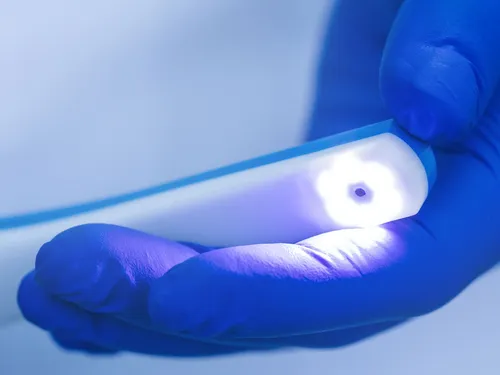
An intraoral camera is a small, pen-sized device with a bright light that takes high-resolution photos and live video of your teeth and gums. It is used to show color, texture, and hard-to-see details during a routine exam.
This digital dental imaging tool helps with early detection of cracked enamel, worn fillings, surface decay, gum inflammation, and plaque or tartar buildup.
How Intraoral Cameras Help You
- Early Identification Of Problems Before They Worsen.
- Visual, Chairside Patient Education So You Can Follow Along In Real Time.
- Accurate Records To Monitor Changes Over Time.
- More Precise Treatment Planning And Case Discussions.
- Noninvasive Imaging With No Radiation.
- Comfortable Use Even In Small Or Sensitive Areas.
The Intraoral Camera Process
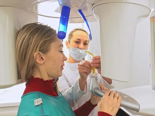
The process is quick and comfortable. A disposable barrier covers the camera for infection control. dentist or clinical team gently moves the small wand across your teeth and gums. Images appear on a monitor immediately.
Most image sets take only a few minutes. Photos are saved to your secure dental record and can be compared at future visits to track healing or wear.
What To Expect During Your Visit
- Before: Brush as usual and share any sensitivity areas with the team.
- During: Watch images live on the screen and ask questions as they arise.
- After: Review key photos and discuss options such as preventive care, fillings, crowns, implants, or denture relines.
Your Options, Supported By Visuals
Intraoral photos make it easier to compare choices like repairing a small cavity with a filling versus placing a crown for a larger fracture. They are useful when planning implants, evaluating gum inflammation, or assessing the fit of dentures. If you seek a second opinion, images provide an objective starting point for discussion.
FAQs
Hours